## Understanding Cystoscopy: A Guide to Medical Testing for Bladder Conditions
### Introduction
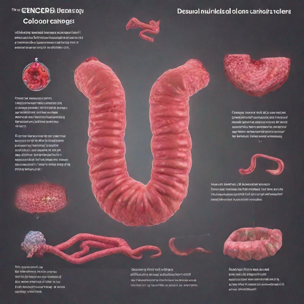
Cystoscopy is a minimally invasive medical test used by urologists** to examine and diagnose **bladder**, **urethra**, and **urinary tract** disorders. It allows doctors to directly visualize the inside of these structures to identify abnormalities and collect samples for further analysis.
### Procedure
During a **cystoscopic** procedure, a thin, flexible instrument called a cystoscope is inserted into the **urethra** and advanced into the **bladder**. The cystoscope is equipped with a **light source** and a **video camera**, which enables the urologist to inspect the inner lining of the **urethra** and **bladder** in real-time and record images for documentation.
In some cases, the urologist may use a **cystoscope with special tools** to:
– Take **biopsies**, small samples of tissue, for examination under a microscope.
– **Remove** small growths or polyps.
– Insert **stents**, small tubes, to keep the urinary tract clear of blockages or damage.
**Two main types of **cystoscopy** are:**
– **Flexible cystoscopy**: Uses a thin, flexible cystoscope that can be maneuvered into different shapes, making it ideal for examining the entire **urinary tract**.
### Diagnosis
**Cystoscopy** is primarily used to diagnose various conditions affecting the **bladder** and **urinary tract**, including:**
– **Urethral strictures**: Narrowing or blockages of **urethra**.
– **Bladder tumors and cancer**: Abnormal growths in or around the bladder’s inner lining.
– **Urinary tract infections (UTIs)**: Infections in any part of **the urinary system**.
– **Incontinence**: Inability or limited ability to empty the bladder.
### Importance
**Cystoscopy** plays an important role in the evaluation of **urinary tract** symptoms and disorders. By providing a direct view of these structures, **cystoscopic** allows urologists:**
– **Accurate diagnosis:** Visualizing the inner lining of **urinary tract organs** helps identify abnormalities, infections, or tumors that may not show up through imaging tests.
– **Early detection of cancer**: **Bladder cancer**, in particular, can be more easily diagnosed and treated at its earliest stages through cystoscopic biopsy.
**Alternatives**
Alternative tests and procedures to **cystoscopy** include:**
– Urine analysis and culture
– Ultrasound
– Computed tomography scan (CT scan).
**However, **cystoscopy** remains the gold standard in diagnosing and assessing conditions affecting the lower **urethral** and **bladder**.
**Preparation**
– Fast for 6-12 hours prior to the procedure to avoid nausea or discomfort during the examination
– Drink plenty of fluids to ensure a clear urine sample.
### Recommendations
Following a cystoscopy, the urologist will discuss the findings with the patient. Based on the results, they may recommend additional tests or treatments, such as:**
– Antibiotics to treat infections
– Surgery to remove tumors or correct structural abnormalities.
– Further imaging studies, including a **Voiding Cystourethrogram (VCUG**) or **Cystogram**.
– **Urodynamics**, special tests to evaluate the function and coordination of **the bladder** and urethra during urination.
– In some instances, a procedure called cystoscopic **laser therapy**, which utilizes laser light, may be performed to remove small bladder tumors or treat overactive bladders.
### Duration
A cystoscopy usually takes about **10 to 30 minutes**. Results are typically available immediately after the procedure.