## Thoracentesis: A Diagnostic and Therapeutic Medical Procedure for Pleural Effusions
### Introduction
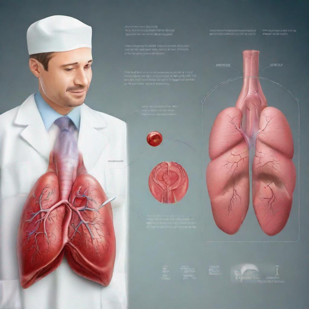
Thoracentesis, also known as chest tap or pleural puncture, is a medical procedure that involves inserting a needle into the pleural space between the lungs and chest wall to withdraw fluid for diagnostic or therapeutic purposes. It is primarily used to evaluate and manage pleural effusions, an abnormal accumulation of fluid in the pleural space.
### Procedure
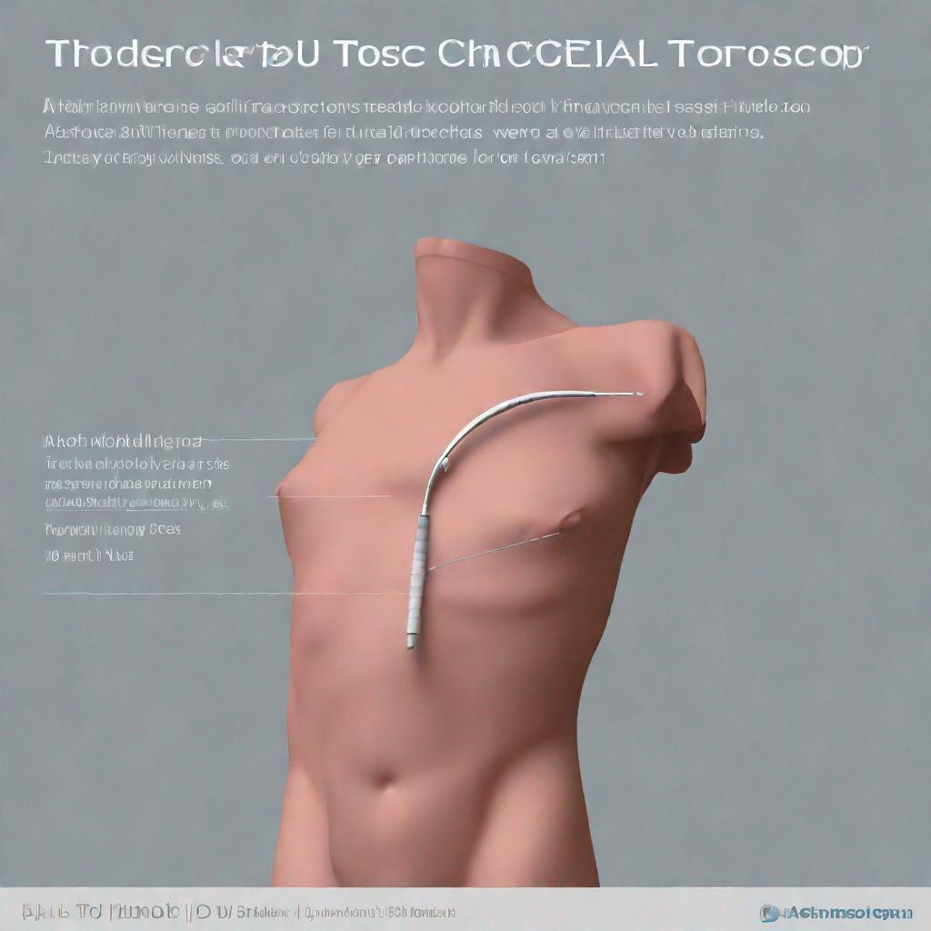
Thoracentesis is typically performed by a pulmonologist, cardiologist, or other healthcare professional experienced in the procedure. Before the procedure, the patient’s chest is cleaned and numbed with a local anesthetic. Then, using ultrasonography or other imaging techniques for guidance, the healthcare provider inserts a small needle into the pleural space. The fluid is then withdrawn into a syringe or collection container.
### Diagnosis
Thoracentesis can aid in the diagnosis of various conditions and diseases that cause pleural effusions, including:
* Heart failure
* Liver failure
* Kidney failure
* Pneumonia
* Pleuritis
* Empyema
* Tuberculosis
* Malignancy
* Cirrhosis
The analysis of the pleural fluid can reveal its color, viscosity, cell count, culture results, and other characteristics that provide clues to the underlying cause of the effusion.
### Importance
Thoracentesis is an important procedure because it allows healthcare providers to:
* Diagnose and differentiate between different types of pleural effusions
* Determine the cause of the effusion
* Relieve symptoms of dyspnea and chest pain by draining excess fluid
* Obtain samples for further testing, such as culture or cytology
* Administer therapeutic agents into the pleural space, such as antibiotics or chemotherapy
### Alternatives
In some cases, alternative tests or procedures may be used to evaluate pleural effusions instead of thoracentesis. These alternatives include:
* Chest radiograph
* Echocardiography
* Computed tomography (CT) scan
* Magnetic resonance imaging (MRI) scan
### Preparation
For thoracentesis, patients are typically asked to do the following:
* Fast for a few hours before the procedure
* Remove any jewelry or clothing that may interfere with the procedure
* Inform the healthcare provider of any medications or allergies
### Duration
Thoracentesis typically takes between 15 and 30 minutes to complete. Patients may experience a slight discomfort during the procedure, but it is usually well-tolerated. Most results are available within 24-48 hours.
### Recommendations
Following thoracentesis, patients may be advised to:
* Rest for a few hours
* Avoid strenuous activity
* Monitor for any symptoms of infection or complications, such as fever or chest pain
* Consider additional tests or procedures, such as a chest CT scan or pericardiocentesis, if necessary
### Related Procedures / Tests
Other related procedures or tests that may be used in conjunction with or following thoracentesis include:
* **Chest tube:** Insertion of a flexible tube into the pleural space to drain larger amounts of fluid or air
* **Chest radiograph:** X-ray imaging of the chest to assess the location and extent of the effusion
* **Echocardiography:** Ultrasound imaging of the heart to rule out cardiac causes of the effusion
* **Computed tomography (CT) scan:** Advanced imaging technique to visualize the pleural space and underlying structures
* **Magnetic resonance imaging (MRI) scan:** Detailed imaging technique to assess the pleural space and surrounding tissues
**Keywords:** Thoracentesis, Pleural effusion, Pleural fluid, Chest tube, Chest tap, Pleurocentesis, Pericardiocentesis, Paracentesis, Ascitic fluid, Pericardial effusion


