
## Optical Coherence Tomography (OCT): A Comprehensive Guide
**Introduction**
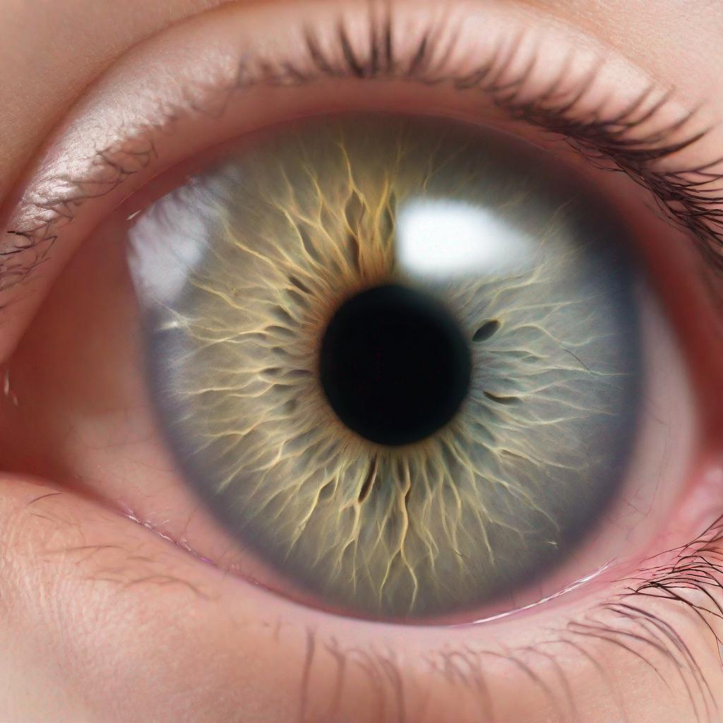
Optical coherence tomography (OCT) is a non-invasive imaging test that uses light waves to create a highly detailed cross-sectional view of the retina, the light-sensitive tissue at the back of the eye. This optical biopsy provides valuable information for diagnosing and monitoring various eye conditions and diseases.
**Procedure**
OCT is typically performed by an ophthalmologist or optometrist. During the test, you will be seated comfortably with your head resting on a chinrest. A small light source is positioned in front of your eye, and harmless light waves are emitted into the eye. The reflected light is captured and analyzed by an OCT machine to create detailed images. The entire procedure is painless and non-invasive.
**Diagnosis**
OCT can identify and evaluate various conditions and diseases, including:
* **Age-related macular degeneration (AMD)**
* **Diabetic retinopathy**
* **Glaucoma**
* **Retinal detachment**
* **Macular hole**
* **Central serous chorioretinopathy (CSC)**
* **Retinal vein occlusion**
* **Diabetic macular edema (DME)**
* **Epiretinal membrane**
**Importance**
OCT is a vital diagnostic tool in ophthalmology due to its ability to:
* **Detect and evaluate early signs of disease** before symptoms appear
* **Monitor and track disease progression** over time
* **Identify potential complications** and guide treatment decisions
* **Assess treatment response and effectiveness**
**Alternatives**
Alternative tests for evaluating the retina include:
* **Fundus photography:** Captures images of the back of the eye
* **Fluorescein angiography:** Uses a special dye to highlight blood vessels
* **Indocyanine green angiography:** Uses a different dye to visualize the choroid
* **Ultrasound biomicroscopy (UBM):** Uses sound waves to create images of the eye structures
**Preparation**
No special preparation is required for an OCT. However, you should inform your doctor if you have any eye conditions or are taking any medications.
**Duration**
OCT typically takes around 10-15 minutes.
**Results**
Results are usually available immediately and interpreted by your doctor. They will discuss the findings with you and recommend any necessary treatment or further testing.
**Recommendations**
Depending on the results of your OCT, your doctor may recommend additional tests or procedures, such as:
* Fundus photography
* Visual field testing
* OCT angiography (OCTA)
* Electroretinography (ERG)