## Dilated Fundus Examination: A Comprehensive Guide
### Introduction
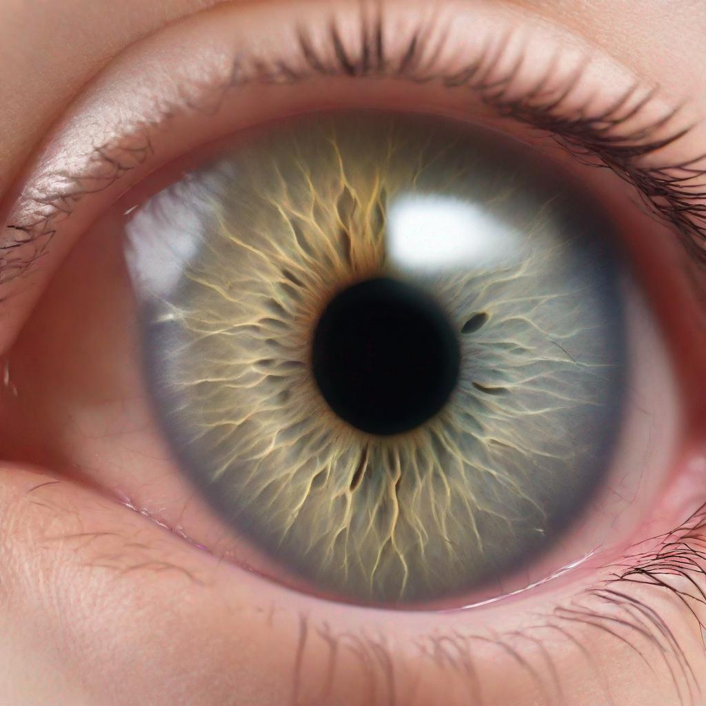
A dilated fundus examination, also known as funduscopy or retinal examination, is a medical test that involves examining the back of the eye, including the retina, macula, optic nerve, and other structures. It is a vital tool in diagnosing and monitoring a wide range of eye conditions and diseases.
### Procedure
**How is the Test Performed?**
1. **Pupil Dilation:** Your doctor will dilate your pupils using eye drops that widen the pupils. This allows for a better view of the back of the eye.
2. **Examination:** Your doctor will use an ophthalmoscope, a lighted instrument, to examine the retina and other structures. The ophthalmoscope may be held up to your eye or placed on your forehead.
3. **Magnification:** The ophthalmoscope has various lenses that magnify the structures of the eye, allowing your doctor to see them in detail.
**Tools Used**
– Ophthalmoscope
– Mydriatic eye drops (for pupil dilation)
– Slit lamp (optional)
**Who Performs the Test?**
Typically, ophthalmologists (eye doctors) or optometrists perform dilated fundus examinations.
### Diagnosis
**What Conditions and Diseases Can be Identified?**
A dilated fundus examination can reveal a variety of eye conditions and diseases, including:
– **Retinal diseases:** Age-related macular degeneration (AMD), diabetic retinopathy, retinal detachment, and macular holes
– **Optic nerve diseases:** Glaucoma, optic neuritis, and optic nerve atrophy
– **Vascular diseases:** Hypertensive retinopathy, retinal vascular occlusions, and diabetic retinopathy
– **Other conditions:** Tumors, infections, and uveitis
### Importance
A dilated fundus examination is crucial for diagnosing and monitoring eye conditions that can lead to vision loss. It allows doctors to:
– Detect early signs of eye diseases
– Monitor the progression of existing conditions
– Assess the effectiveness of treatments
– Make informed decisions about patient care
### Alternatives
There are other tests and procedures that can be used to examine the back of the eye, such as:
– **Fundus photography:** Captures images of the retina
– **Fundus fluorescein angiography (FFA):** Injects dye into the bloodstream and photographs the retina to assess blood flow
– **Spectral domain optical coherence tomography (SD-OCT):** Uses light to create cross-sectional images of the retina
### Preparation
Before the test, inform your doctor about any medications you are taking, especially blood thinners. Avoid wearing eye makeup or contact lenses. Bring a pair of sunglasses to wear after the test, as your pupils will be dilated.
### Duration
The test usually takes about 15-30 minutes. The dilation effects may last for several hours, affecting your vision.
### Recommendations
Following a dilated fundus examination, your doctor may recommend additional tests or procedures based on the findings:
– **Dilated pupil examination:** To further assess the retina and optic nerve
– **Visual field test:** To check for areas of vision loss
– **Optical coherence tomography (OCT):** To create detailed images of the retina
– **Fluorescein angiography (FA):** To evaluate blood flow in the retina