## Cerebral Angiography: A Comprehensive Guide
### Introduction
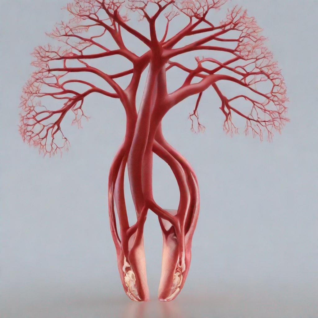
Cerebral angiography is a medical imaging test that provides detailed images of the blood vessels in the brain. It is used to diagnose and treat various conditions affecting the brain’s blood supply.
### Procedure
The test involves injecting a dye into the arteries of the brain through a catheter inserted into the femoral artery (transfemoral angiography) or the brachial artery (transbrachial angiography). The dye travels through the arteries, and X-ray images are taken to visualize the blood vessels and identify any abnormalities.
The procedure is typically performed by a team of interventional radiologists or neurologists. It may take several hours and requires the patient to remain still during the imaging.
### Diagnosis
Cerebral angiography is used to diagnose and assess the severity of several conditions, including:
– **Cerebral aneurysm:** A weakened area of an artery in the brain that can rupture and cause a hemorrhage.
– **Arteriovenous malformation (AVM):** An abnormal connection between arteries and veins in the brain.
– **Carotid artery stenosis:** Narrowing of the carotid arteries, which supply blood to the brain.
– **Stroke:** A brain injury caused by a blockage or rupture of a blood vessel in the brain.
– **Brain tumor:** A growth of abnormal cells in the brain.
– **Vascular dementia:** A progressive decline in cognitive function due to impaired blood flow to the brain.
### Importance
Cerebral angiography is a valuable diagnostic tool because it provides detailed images of the brain’s blood vessels, allowing doctors to:
– Accurately diagnose vascular conditions.
– Determine the location and severity of abnormalities.
– Plan appropriate treatment strategies.
– Monitor the effectiveness of treatment over time.
### Alternatives
Alternative tests to cerebral angiography include:
– **Computed tomography angiography (CTA):** A non-invasive imaging test that uses X-rays and a dye to create images of the brain’s blood vessels.
– **Magnetic resonance angiography (MRA):** A non-invasive imaging test that uses magnetic fields and radio waves to create images of the brain’s blood vessels.
### Preparation
Before the test, the patient may be asked to:
– Fast for several hours.
– Adjust medications as directed by the doctor.
– Avoid caffeine and smoking.
– Inform the doctor of any allergies or medical conditions.
### Duration
The test usually takes 1-3 hours. The patient may need to stay in the hospital for a few hours after the procedure for observation.
### Recommendations
Following cerebral angiography, the doctor may recommend other tests or procedures, such as:
– **Carotid endarterectomy:** Surgery to remove plaque from the carotid arteries.
– **Stent placement:** Implantation of a mesh tube to open narrowed arteries.
– **Coil embolization:** Treatment to block aneurysms or AVMs.
– **Radiation therapy:** Treatment to shrink brain tumors.