## Aortography: A Comprehensive Guide
Introduction
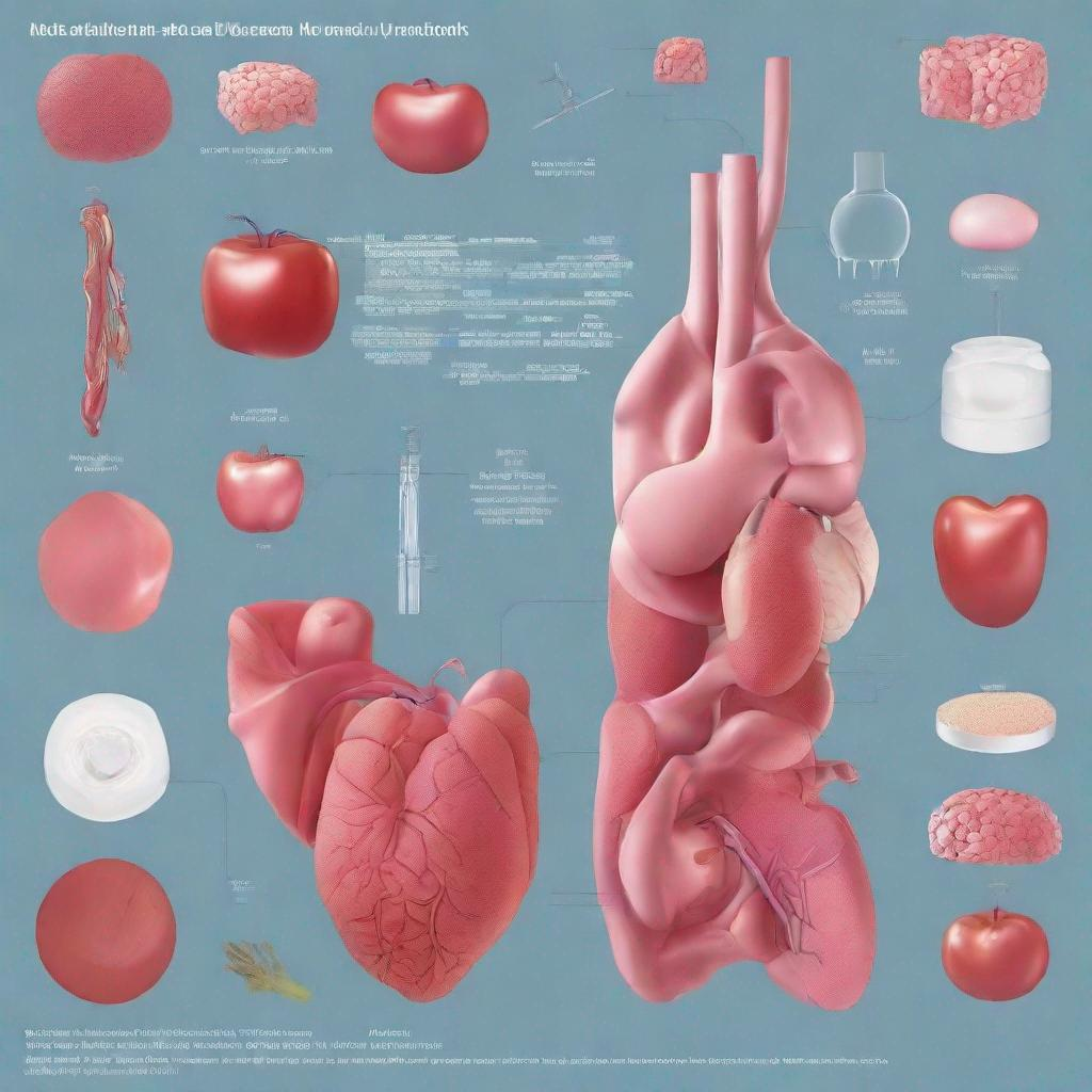
Aortography is a medical imaging test that allows doctors to visualize the aorta, the largest artery in the body, and its branches. It is used to diagnose and evaluate various conditions and diseases affecting the aorta and its surrounding structures.
Procedure
Aortography is typically performed by an interventional radiologist or a vascular surgeon. The procedure involves:
* Injecting a contrast dye into a peripheral artery, usually in the groin or arm.
* Using fluoroscopy, a real-time X-ray imaging technique, to guide the dye as it travels through the aorta.
* Taking multiple X-ray images to capture the blood flow and the structure of the aorta.
Diagnosis
Aortography can help diagnose and evaluate:
* **Abdominal Aortic Aneurysm (AAA):** A bulging or weakened area in the abdominal aorta.
* **Aortic Stenosis:** A narrowing of the aortic valve, which can restrict blood flow.
* **Aortic Dissection:** A tear in the inner lining of the aorta.
* **Arteriovenous Malformation (AVM):** An abnormal connection between an artery and a vein.
* **Coarctation of Aorta:** A narrowing of the aorta that can block blood flow to the lower extremities.
Importance
Aortography is a valuable diagnostic tool for evaluating the aorta and its branches. It provides detailed images that can help doctors:
* Determine the size, shape, and location of an aneurysm or dissection.
* Assess the severity of aortic stenosis.
* Locate and characterize AVMs.
* Plan treatment strategies for these conditions.
Alternatives
Alternative imaging tests that can be used to evaluate the aorta include:
* **Angiography:** Similar to aortography, but uses a catheter inserted into the aorta to inject the contrast dye.
* **Computed Tomography Angiography (CTA):** A non-invasive imaging technique that uses X-rays and computer processing to create detailed images of the aorta.
* **Magnetic Resonance Angiography (MRA):** A non-invasive imaging technique that uses magnetic fields and radio waves to create images of the aorta.
Preparation
Prior to the test, patients may need to:
* Fast for several hours before the procedure.
* Stop taking certain medications, such as blood thinners.
* Inform their doctor about any allergies or other medical conditions.
Duration
Aortography typically takes about 30-60 minutes to perform. The results may be available immediately or within a few days.
Recommendations
Following aortography, your doctor may recommend other tests or procedures, such as:
* **Transesophageal Echocardiography:** An ultrasound exam that provides detailed images of the heart and aorta.
* **Stent Placement:** A surgical procedure to insert a device that can open up a narrowed or blocked aorta.
* **Aortic Valve Replacement:** A surgical procedure to replace a damaged or dysfunctional aortic valve.